


For decades, open surgical repair was the only available method to treat CWDs. 20-30 years ago, elasticity and flexibility of the ventral chest wall were identified as an important factor for successful treatment of CWDs. Consequently, non-surgical measures such as vacuum bell therapy for conservative treatment of PE and customized bracing systems for conservative treatment of PC were established. In 1998, D. Nuss described a minimally invasive procedure for surgical repair of Pectus excavatum (PE), and a few years later H. Abramson reported on his experience with the “reversed Nuss technique” for surgical repair of Pectus carinatum (PC). Both procedures are performed with increasing frequency worldwide.
Nowadays information on new therapeutic modalities circulate not only among pectus surgeons and paediatricians, but also rapidly among patients. Pectus surgeons have to be aware that assessment of a CWD patient has to be more differentiated. A CWD has to be considered as a progressive deformity, since we usually observe a worsening of the deformity during periods of rapid growth in the majority of CWD patients. Self-perception is age dependent and completely different between pediatric and adolescent patients. Analysing the increasing number of studies reporting on conservative treatment of CWDs as well as our own group of patients who visited a busy outpatient clinic in a small country within the last 15 to 20 years, we have to realize that the majority of patients asked for non-surgical treatment of their CWD.
This overview summarizes current concepts and new trends in surgical and non-surgical treatment methods of congenital and acquired CWDs.
Introduction
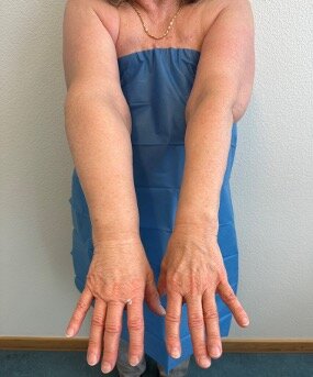
Pectus Excavatum (PE) and Pectus carinatum (PC) represent the most common chest wall deformities (CWDs), occurring in approximately 1 in every 300-400 births, and showing a male predominance (approx. 4:1 ratio). A recent published study analysing more than 600 artefacts from Ancient Egypt, showed that CWDs were already displayed in reliefs from Ancient Egypt dating back to circa 2400 BC.1 In many CWD patients, asymmetry of the ventral chest wall, unilateral and/or bilateral costal flaring as well as concomitant scoliosis and/or kyphosis of the spine may be noticed. Some patients may present with combined CWD such as pectus arcuatum (PA) or so called Currarino-Silverman syndrome (Figure 1).

Figure 1: 12y old male patient with Currarino-Silverman syndrome
For decades and until turn of the millennium resp., commonly held beliefs include the opinion that CWD, in particular PE, is “only a cosmetic problem”, and patients do not suffer from relevant clinical symptoms. Furthermore, previously used procedures to correct CWDs were largely based on open surgical techniques, such as the Ravitch procedure and its modifications.2–6 In 1992 Haje from Brazil reported first time on successful conservative brace therapy in PC patients, incorporating elasticity and flexibility of the ventral chest wall as an important factor for successful treatment.7 Donald Nuss introduced 1998 the technique of minimally invasive repair of Pextus excavatum (MIRPE) to avoid several operative features of the modified Ravitch repair procedure.8 In 2005, H. Abramson reported on his preliminary experience with a minimally invasive technique using a subcutaneous pectus bar to correct PC (MIRPC), the so-called reversed Nuss-procedure.9 The introduction of these minimally invasive techniques and subsequent modifications, new therapeutic modalities such as more sophisticated bracing systems for conservative treatment of PC, the vacuum bell therapy (VBT) for conservative treatment of PE as well as an increasing interest and patient’s introspection have changed the view on the treatment of CWDs within the last 25 years.10 New information circulate not only among surgeons and paediatricians, but also rapidly among patients. Furthermore, patients who refused operative treatment by previously available procedures due to several reasons, now appear at the outpatient clinic and request to be considered for the new, non- surgical methods.10
Today, pectus surgeons are aware that assessment of a CWD patient has to be more differentiated and sophisticated. Pediatric patients with CWD will usually have less symptoms compared to adolescent and/or adult CWD patients. Involvement in physical activity is an important aspect and will usually change over time. A CWD has to be considered as a progressive deformity. Therefore, we observe a worsening of the deformity during periods of rapid growth in the majority of CWD patients. Self-perception is age dependent and completely different between pediatric and adolescent patients.
This overview summarizes current concepts and new trends in surgical and non-surgical treatment methods of congenital and acquired CWDs.
Surgical Repair of pectus excavatum - MIRPE
The MIRPE technique was introduced in 1998 8, is today well established and represents the worldwide used “gold-standard” for surgical repair of PE in adolescent PE patients. Several reports on risks and near fatal complications during MIRPE have helped pectus surgeons to develop technical modifications.11 Notably the use of routine unilateral and/or bilateral thoracoscopy and intraoperative sternal elevation during MIPRE have resulted in increased safety and a lower rate of overall complications.12 MIRPE should be done under careful visualization by thoracoscopy, always having the tip of the introducer under direct visual control during retrosternal dissection. Sternal elevation is important to improve safety of MIRPE.12–14 Proper bar placement is considered as a crucial precondition and mandatory for safe pectus bar removal.13
Age at Time of MIRPE
In contrast to the first report by D. Nuss where the majority of operated patients were under the age of 10 years and even if some pectus surgeons perform MIPRE in pediatric patients, there is general consent today to perform MIRPE at the age of 13 to 16 years.15 This covers the period when the deformity may become more severe, more complex and more asymmetric. In particular in female patients, bilateral skin incisions may be done at the insertion of the breast, becoming later nearby invisible (Figure 2).

Figure 2: 13y old female patient with PE before and after MIRPE

The implant should remain for approx. 3 years.13,16 Performing MIRPE in pediatric patients, you have to be aware of an increased risk of recurrence after pectus bar removal due to pubertal growth spurt dependent influence. Performing MIRPE in adult patients, other age related aspects have to be considered.13,17
Number and Position of Pectus bar(s)
Not only the number of pectus bars but also positioning has changed over time. Initially and for many years, placement of one pectus bar including one lateral stabilizer on the non-dominant side was routine standard procedure.15,18 Modifications included the use of a second stabilizer, a second or a third bar, with or without stabilizers.15,19 In particular in patients with a severe PE deformity such as Grand Canyon type, use of a second bar or in even a third or fourth bar has to be considered.20 The so-called cross-bar technique was established to correct a wider range of PE deformities, especially at the lower part of the depression.20,21 HJ Park introduced the total chest wall remodeling to correct complex combined deformities.20 Furthermore, Bellia-Munzon et al. reported on their preliminary experience with 3D reconstruction and printing of customized pectus bars developed by computer-aided design and digital image technology.22
Sternal elevation during MIRPE
Within the last decade, an increasing number of authors report on the routine use of sternal elevation during MIRPE to improve the safety of the procedure. In particular in adult PE patients, stiffness and rigidity of the chest wall may hamper the retrosternal dissection. Sternal elevation guarantees a proper visualization of the retrosternal area. Elevation methods include the crane technique introduced by HJ Park, an additional subxiphoid incision, the use of various handhold devices (e. g. Langenbeck retractor, Wolkmann bone hook, etc.) as well as the intraoperative use of the vacuum bell. 12,14 The safety of MIRPE has improved clearly as there has no near-fatal incident reported anymore since sternal elevation was applied intraoperatively. In our unit, we have excellent experience with the intraoperative use of the vacuum bell for approx. 20 years.12
Cryoablation for pain management
MIRPE is associated with significant pain, and efforts to control pain impact resource utilization. Efficient pain management is a crucial and mandatory measure to ensure a good outcome and to reduce hospital stay. Vice versa, poor pain control not only increases hospital stay, but also can raise opioid consumption, limit mobility and/or favour readmissions. Traditional pain control included patient controlled analgesia or epidural. Within the last couple of years, an increasing number of authors reported on their experience with cryoablation for pain management.23–25 The application can be performed as ultrasound-guided percutaneous bilateral cryoanalgesia one or two days before MIRPE,26 or as bilateral thoracoscopic procedure within the same anesthesia prior to MIRPE. A recent published study reported on the benefits of additional erector spinae plane block before cryoanalgesia during MIRPE.27 However, applicants have to be aware of possible side effects such as neuropathic pain and/or long-term sensory function impairment.23 In our unit, we use bilateral thoracoscopic cryoablation for more than one year with excellent results. Cryoablation reduces median total opioid requirement as well as length of hospital stay.
Conservative Treatment of Pectus Excavatum
Especially pediatric patients present often with a mild degree of PE, which is stable during childhood, even if the deformity is noticeable at birth. An increase of PE severity is usually observed in nearby all patients during pubertal growth spurt. Of course, in many PE patients the degree of pectus deformity does not immediately warrant surgery. Nonetheless, some PE patients may benefit from some type of non-surgical treatment. Pain associated with postoperative recovery and the risk of imperfect results after surgery represent important aspects, which may affect patients and parents’ decision about type of treatment. The introduction of vacuum bell therapy (VBT) for conservative treatment of PE has made this alternative therapy a focus of interest of patients, their parents as well as surgeons.
VBT represents today a well-established complement in the treatment of PE. Although described already more than 100 years ago28, the routine use of VBT could not be established for a long time due to different reasons. First after turn of the millennium, pilot studies reported encouraging and promising results.29,30 CT-scans showed that the device lifted the sternum and ribs immediately.29 Afterwards, several studies reported on an increasing number of patients31,32, and the first systematic retrospective study summarizing the results of 133 patients was published in 2011.33 Latest reports confirmed the efficacy of VBT in carefully selected patients.34–39 In our largest series including more than 500 patients, approximately 80% of PE patients applied for VBT whereas 20% underwent surgical repair.37 This is in concordance with other larger PE patients series.10 Identifying relevant variables for a good to excellent outcome, a recent published study could corroborate our experience. Obermeyer et al. reported on an excellent outcome for patients ≤ 11 years, chest wall depth ≤ 1.5cm, chest wall flexibility, and VB use over 12 consecutive months.39 Today, VBT has been established as an additional useful tool in specific treatment of patients suffering from PE, providing good to excellent results (figure 3).

Figure 3: 13y old male patient with PE before and after VBT

VBT represents no contradiction, but a complement in the specific treatment of PE patients, applicable to a significant majority of PE patients. Furthermore, VBT may allow some patients with PE to avoid surgery. In our chest wall unit, we offer VBT for pediatric, adolescent as well as adult PE patients for more than 20 years. However, careful patients’ selection is mandatory to avoid any severe disappointments.
Treatment of Pectus Carinatum
Similar to PE, the natural history of PC is of a mild defect seen in infancy, which is stable during childhood. Again similar to PE and PA, the deformity usually shows a significant worsening during pubertal growth spurt. Most patients with PC show mild or no relevant clinical symptoms. Significant physical or cardiopulmonary symptoms are usually not observed. The most frequently reported symptoms are tenderness, bone pain or mild exercise intolerance. However, the majority of PC patients have a disturbed body image, and the patient`s introspection may be impaired.40 As a consequence, reduced quality of life is the key point for the majority of PC patients and the most relevant motivation to seek medical advice. To the best of our knowledge, there are no objective parameters to indicate any type of treatment for PC patients as we know it from PE patients. Dependent on the patient`s age and the severity of the deformity, current treatment options include observation, bracing or surgical repair. Considering that the anterior chest wall is still compliant during puberty and that it permits remodelling by external compression, Haje from Brazil inaugurated conservative brace therapy and reported his experience first time in 1992.7 Over the last 25 years, bracing has gained significant popularity among surgeons and patients, and became the first line treatment for PC, applying different models of customized external braces.7,41–47 Martinez-Ferro and Fraire developed a dynamic compression system (DCS), enabling the surgeon to measure the pressure of initial correction (PIC) to correct PC as well as the pressure of treatment (POT) which may be used during application of the DCS.48–50 The DCS scales the pressure measurement during therapy (POT), permits prediction of treatment duration and prognosis, permits in-situ outpatient clinic adjustments and avoids patient manipulation.49,50 Today, the effectiveness of DCS treatment could be confirmed by numerous studies.40,51–55 In our chest wall unit, we offer orthotic bracing using orthotic braces, either static carbon brace or DCS, both with good to excellent results. As confirmed by different authors, we consider patients under the age of 14-15 years with symmetric PC and high motivation toward correction as the best candidates for this treatment method.
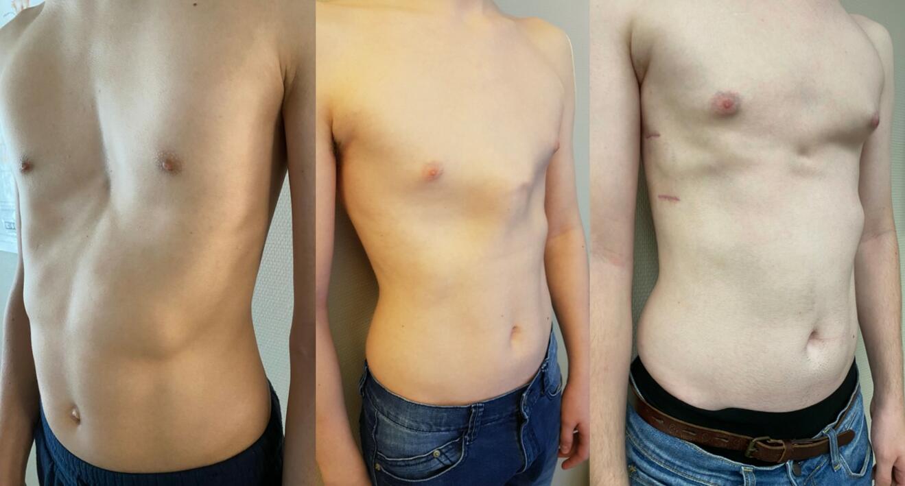
Only a minority of PC patients needs surgical repair. Frey et al. reported surgical repair in 17% of PC patients.41 In our study group, only 3% (2/68 PC patients) underwent surgical repair.40 The MIRPC procedure performed by experienced surgeons provides excellent results, but side effects and complications have to be noticed.19,56–58 In case of a complex combined deformity, total chest wall remodeling inaugurated by HJ Park may be indicated to guarantee a good to excellent result (figure 4).20

Figure 4: 17y old male patient with complex combined deformity before and after chest wall remodelling

Summarizing these results, orthotic bracing is considered as the most important and effective treatment modality for PC patients. This may be confirmed by the fact that we found much more studies on bracing than on surgical repair in the current literature concerning specific treatment of PC. Furthermore, the majority of PC patients in our chest wall unit is treated with orthotic bracing.
Conclusion
Specific treatment is not necessary in every patient with a CWD, but observation is mandatory, especially in pediatric and adolescent patients. Dependent on patient’s age and severity of the CWD, non-surgical treatment might be the first step of therapy. In PC patients, orthotic bracing represents an effective treatment modality and should be considered as first choice treatment. Only a minority of PC patients needs surgical repair. VBT seems to be safe and has been established as an additional useful tool in specific treatment of PE patients, in particular in pediatric and young adolescent patients. For surgical repair of PE, MIRPE is today well established and represents the worldwide used “gold-standard”, especially in adolescent PE patients. The routine use of thoracoscopy as well as intraoperative SET is considered as mandatory to improve safety of the procedure and to avoid near-fatal complications like cardiac injury, respectively. Patients and parents may appreciate if the pectus surgeon may offer all different treatment modalities.
- Bialas AJ, Kaczmarski J, Kozak J, Kempinska-Miroslawska B. Pectus excavatum in relief from Ancient Egypt (dating back to circa 2400 BC). Interact Cardiovasc Thorac Surg. 2015;20(4):556-557. doi:10.1093/icvts/ivu440
- Ravitch MM. The operative treatment of pectus excavatum. Ann Surg. 1949;129:429-444.
- Ravitch MM. Operative Correction of Pectus Carinatum (Pigeon Breast). Ann Surg . 1960;151:705-715.
- Shamberger RC, Welch KJ. Surgical Correction of Pectus Carinatum. J Pediatr Surg. 1987;22:48-53.
- Robicsek F. Surgical treatment of pectus carinatum. Chest Surg Clin N Am. Published online 2000:357-376.
- Fonkalsrud EW. 912 Open pectus excavatum repairs: Changing trends, lessons learned: One surgeon’s experience. World J Surg. 2009;33(2):180-190. doi:10.1007/s00268-008-9793-4
- Haie S, Bowen J. Preliminary results of orthotic treatment of pectus deformities in children and adolescents. J Pediatr Orthop. 1992;12:795-800.
- Nuss D, Kelly R, Croitoru D, Katz M, J Pediatr Surg ND. A 10-Year Review of a Minimally Invasive Technique for the Correction of Pectus Excava-tum. J Pediatr Surg. 1998;33:545-552.
- Abramson H. A minimally invasive technique to repair pectus carinatum. Preliminary report. Arch Bronchoneumol. 2005;41:349-351.
- Häcker FM. Nonsurgical Treatment of Chest Wall Deformities: Contradiction or Complement? European Journal of Pediatric Surgery. 2018;28(4):369-372. doi:10.1055/s-0038-1668128
- Becmeur F, Ferreira C, Haecker F, Schneider A, Lacreuse I. Pectus excavatum repair according to Nuss: is it safe to place a retrosternal bar by a transpleural approach under thoracoscopic vision? Journal of Laparoendoscopic & Advanced Surgical Techniques. 2011;A8:757-761.
- Haecker FM, Krebs T, Kocher GJ, Schmid RA, Sesia SB. Sternal elevation techniques during the minimally invasive repair of pectus excavatum. Interact Cardiovasc Thorac Surg. 2019;29(4). doi:10.1093/icvts/ivz142
- Frank-Martin Haecker, Andre Hebra, Marcelo Martinez Ferro. Pectus bar removal - why, when, where and how. J Pediatr Surg. 2021;56(3):540-544.
- Obermeyer RJ, Goretsky MJ, Kelly RE, et al. Selective use of sternal elevation before substernal dissection in more than 2000 Nuss repairs at a single institution. J Pediatr Surg. 2021;56(4):649-654. doi:10.1016/j.jpedsurg.2020.07.005
- Kelly RE, Goretsky MJ, Obermeyer R, et al. Twenty-one years of experience with minimally invasive repair of pectus excavatum by the Nuss procedure in 1215 patients. Ann Surg. 2010;252(6):1072-1081. doi:10.1097/SLA.0b013e3181effdce
- Nuss D, Obermeyer RJ, Kelly RE. Pectus excavatum from a pediatric surgeon’s perspective. Ann Cardiothorac Surg. 2016;5(5):493-500. doi:10.21037/acs.2016.06.04
- Zoeller GK, Zallen GS, Glick PL. Cardiopulmonary resuscitation in patients with a Nuss bar - A case report and review of the literature. J Pediatr Surg. 2005;40(11):1788-1791. doi:10.1016/j.jpedsurg.2005.07.036
- Kelly RE. Pectus excavatum: historical background, clinical picture, preoperative evaluation and criteria for operation. Semin Pediatr Surg. 2008;17(3):181-193. doi:10.1053/j.sempedsurg.2008.03.002
- Wang W, Chen C. Minimally invasive repair of pectus carinatum: bar number and technique. . Ann Thorac Surg. 2018;12.
- Hyun K, Park HJ. The Cross-Bar Technique for Pectus Excavatum Repair: A Key Element for Remodeling of the Entire Chest Wall. European Journal of Pediatric Surgery. 2022;33(4):310-318. doi:10.1055/a-1897-7202
- Haecker FM, Krebs TF, Kleitsch KU. To Cross or Not to Cross: The Cross-Bar Technique to Correct Pectus Excavatum With “Costal Flaring.” Annals of Thoracic Surgery Short Reports. 2023;1(1):107-110. doi:10.1016/j.atssr.2022.10.019
- Bellia-Munzon G, Martinez J, Toselli L, et al. From bench to bedside: 3D reconstruction and printing as a valuable tool for the chest wall surgeon. J Pediatr Surg. 2020;55(12):2703-2709. doi:10.1016/j.jpedsurg.2020.07.010
- Eldredge RS, McMahon L. Intercostal nerve cryoablation therapy for the repair of pectus excavatum: a systematic review. Front Surg. 2023;10. doi:10.3389/fsurg.2023.1235120
- Linton SC, Tian Y, Zeineddin S, et al. Intercostal Nerve Cryoablation Reduces Opioid Use and Length of Stay Without Increasing Adverse Events : A Retrospective Cohort Study of 5442 Patients Undergoing Surgical Correction of Pectus Excavatum. Ann Surg. 2024;279(4):699-704. doi:10.1097/SLA.0000000000006113
- Toselli L, Gigena C, Bellia-Munzon G, Sanjurjo D, Vallee M, Martinez-Ferro M. Lessons Learned after 176 Patients Treated with a Standardized Procedure of Thoracoscopic Cryoanalgesia during Minimally Invasive Repair of Pectus Excavatum. J Pediatr Surg. 2024;59(3):372-378. doi:10.1016/j.jpedsurg.2023.10.047
- Velayos M, Alonso M, Delgado-Miguel C, et al. Percutaneous Cryoanalgesia: A New Strategy for Pain Management in Pectus Excavatum Surgery. European Journal of Pediatric Surgery. 2022;32(1):73-79. doi:10.1055/s-0041-1740555
- Zacha S, Jarosz K, Kokot K, et al. Benefits of the Erector Spinae Plane Block before Cryoanalgesia in Children Undergoing Surgery for Funnel Chest Deformity. J Pers Med. 2023;13(12). doi:10.3390/jpm13121696
- Lange F. Thoraxdeformitäten. In: Pfaundler M, Schlossmann A, eds. Handbuch Der Kinderheilkunde. Vol 5. FCW Vogel; 1910:157.
- Schier F, Bahr M, Klobe E. The vacuum chest wall lifter: An innovative, nonsurgical addition to the management of pectus excavatum. J Pediatr Surg. 2005;40(3):496-500. doi:10.1016/j.jpedsurg.2004.11.033
- Haecker FM, Mayr J. The vacuum bell for treatment of pectus excavatum: An alternative to surgical correction? European Journal of Cardio-thoracic Surgery. 2006;29(4):557-561. doi:10.1016/j.ejcts.2006.01.025
- Haecker F. Die Saugglocke nach E. Klobe zur konservativen Therapie der Trichterbrust: die Glocke als Alternative zum Bügel? Orthopädische Praxis. 2009;45(4):183-189.
- Haecker FM. The vacuum bell for treatment of pectus excavatum:An effective tool for conservative therapy. Journal of Clinical and Analytical Medicine. 2011;2(1):1-4. doi:10.4328/JCAM.314
- Haecker FM. The vacuum bell for conservative treatment of pectus excavatum: The Basle experience. Pediatr Surg Int. 2011;27(6):623-627. doi:10.1007/s00383-010-2843-7
- Häcker FM, Zuppinger J, Sesia SB. Die konservative Therapie der Tr ichterbrust mittels Vakuumtherapie. Schweizerisches Medizin-Forum. 2014;14(45):842-849. www.trichterbrust.de
- Lopez M, Patoir A, Costes F, Varlet F, Barthelemy JC, Tiffet O. Preliminary study of efficacy of cup suction in the correction of typical pectus excavatum. J Pediatr Surg. 2016;51(1):183-187. doi:10.1016/j.jpedsurg.2015.10.003
- Haecker F, Sesia S. Non-surgical treatment of pectus excavatum. J Visc Surg. 2016;2:63-71.
- Haecker FM, Sesia S. Vacuum bell therapy. Ann Cardiothorac Surg. 2016;5(5):440-449. doi:10.21037/acs.2016.06.06
- Togoro SY, Tedde ML, Eisinger RS, Okumura EM, de Campos JRM, Pêgo-Fernandes PM. The Vacuum Bell device as a sternal lifter: An immediate effect even with a short time use. J Pediatr Surg. 2018;53(3):406-410. doi:10.1016/j.jpedsurg.2017.04.016
- Obermeyer RJ, Cohen NS, Kelly RE, et al. Nonoperative management of pectus excavatum with vacuum bell therapy: A single center study. J Pediatr Surg. 2018;53(6):1221-1225. doi:10.1016/j.jpedsurg.2018.02.088
- Sesia SB, Holland-Cunz S, Häcker FM. Dynamic compression system: An effective nonoperative treatment for pectus carinatum: A single center experience in Basel, Switzerland. European Journal of Pediatric Surgery. 2016;26(6):481-486. doi:10.1055/s-0035-1570758
- Frey AS, Garcia VF, Brown RL, et al. Nonoperative management of pectus carinatum. J Pediatr Surg. 2006;41(1):40-45. doi:10.1016/j.jpedsurg.2005.10.076
- Banever GT, Konefal SH, Gettens K, Moriarty KP. Nonoperative Correction of Pectus Carinatum with Orthotic Bracing. Journal of Laparoendoscopic & Advanced Surgical Techniques. 2006;16(2):164-167.
- Emil S, Laberge JM, Sigalet D, Baird R. Pectus carinatum treatment in Canada: Current practices. In: Journal of Pediatric Surgery. Vol 47. ; 2012:862-866. doi:10.1016/j.jpedsurg.2012.01.035
- Lee RT, Moorman S, Schneider M, Sigalet DL. Bracing is an effective therapy for pectus carinatum: Interim results. J Pediatr Surg. 2013;48(1):184-190. doi:10.1016/j.jpedsurg.2012.10.037
- Colozza S, Bütter A. Bracing in pediatric patients with pectus carinatum is effective and improves quality of life. In: Journal of Pediatric Surgery. Vol 48. ; 2013:1055-1059. doi:10.1016/j.jpedsurg.2013.02.028
- Loff S, Sauter H, Wirth T, Otte R. Highly Efficient Conservative Treatment of Pectus Carinatum in Compliant Patients. European Journal of Pediatric Surgery. 2014;25(5):421-424. doi:10.1055/s-0034-1384648
- Wahba G, Nasr A, Bettolli M. A less intensive bracing protocol for pectus carinatum. J Pediatr Surg. 2017;52(11):1795-1799. doi:10.1016/j.jpedsurg.2017.01.057
- Martinez-Ferro M, Fraire C, Bernard S. Dynamic compression system for the correction of pectus carinatum. Semin Pediatr Surg. 2008;17(3):194-200. doi:10.1053/j.sempedsurg.2008.03.008
- Haecker FM, Martinez-Ferro M. Non-surgical treatment for pectus excavatum and carinatum. In: Chest Wall Deformities and Corrective Procedures. Springer International Publishing; 2015:137-160. doi:10.1007/978-3-319-23968-2_17
- Martinez-Ferro M, Fraire C. Dynamic compressor system for pectus carinatum. In: Saxena A, ed. Chest Wall Deformities. Springer Verlag; 2017:523-542.
- Cohee AS, Lin JR, Frantz FW, Kelly RE. Staged management of pectus carinatum. J Pediatr Surg. 2013;48(2):315-320. doi:10.1016/j.jpedsurg.2012.11.008
- Lopez M, Patoir A, Varlet F, et al. Preliminary study of efficacy of dynamic compression system in the correction of typical pectus carinatum. European Journal of Cardio-thoracic Surgery. 2013;44(5). doi:10.1093/ejcts/ezt425
- Emil S, Sévigny M, Montpetit K, et al. Success and duration of dynamic bracing for pectus carinatum: A four-year prospective study. J Pediatr Surg. 2017;52(1):124-129. doi:10.1016/j.jpedsurg.2016.10.032
- de Beer SA, Gritter M, de Jong JR, van Heurn ELW. The Dynamic Compression Brace for Pectus Carinatum: Intermediate Results in 286 Patients. Annals of Thoracic Surgery. 2017;103(6):1742-1749. doi:10.1016/j.athoracsur.2016.12.019
- Poola AS, Pierce AL, Orrick BA, et al. A Single-Center Experience with Dynamic Compression Bracing for Children with Pectus Carinatum. European Journal of Pediatric Surgery. 2018;28(1):12-17. doi:10.1055/s-0037-1606845
- Pérez D, Cano JR, Quevedo S, López L. New minimally invasive technique for correction of pectus carinatum. European Journal of Cardio-thoracic Surgery. 2011;39(2):271-273. doi:10.1016/j.ejcts.2010.05.024
- Murphy BL, Naik ND, Roskos PL, et al. Minimal cosmetic revision required after minimally invasive pectus repair. Pediatr Surg Int. 2018;34(7):775-780. doi:10.1007/s00383-018-4275-8
- Yuksel M, Lacin T, Ermerak NO, Sirzai EY, Sayan B. Minimally Invasive Repair of Pectus Carinatum. Annals of Thoracic Surgery. 2018;105(3):915-923. doi:10.1016/j.athoracsur.2017.10.003